

How Tianeptine Ethyl Ester Remodels the Central Nervous Signal Transduction Network?
Tianeptine Ethyl Ester is an ethyl esterified derivative of tianeptine, with the molecular formula C₂₃H₂₉ClN₂O₄S and a molecular weight of 465.01 g/mol. Tianeptine itself is a structurally unique atypical antidepressant whose mechanism of action involves the regulation of the glutamatergic system and enhanced serotonin reuptake. However, Tianeptine Ethyl Ester is not the active form of tianeptine. In medicinal chemistry, carboxylic acid drugs are often modified by esterification to improve lipid solubility or as prodrugs. This ethyl ester derivative itself is not directly used as a therapeutic agent in the mainstream international pharmaceutical market. Its core value lies more in two aspects: as a key intermediate or process impurity in the synthesis of tianeptine, and as a reference standard for the quantitative detection of tianeptine content in high-performance liquid chromatography (HPLC).
🧪 Ester modification alters transmembrane transport and intracellular transformation patterns
Tianeptine Ethyl Ester retains the characteristic dibenzothiaza tricyclic skeleton of tianeptine. The carboxyl group at the end of the seven-carbon aliphatic side chain is sealed by an ethyl ester group, eliminating the strong polarity of the carboxylic acid group. This results in a significant shift in the overall lipophilic partition coefficient, enhancing lipophilicity. Tianeptine Ethyl Ester disperses uniformly in physiological buffers and cell culture media without significant crystal precipitation. In high-throughput neuronal drug delivery screening systems, it ensures consistent initial exposure concentrations across different culture wells, reducing data discrepancies caused by solubility differences in parallel samples. The tricyclic conjugated skeleton exhibits strong chemical stability; even under light-protected, low-temperature storage, 37°C cell incubation, and repeated freeze-thaw cycles, the parent nucleus is not prone to oxidation and ring-opening. Stock solutions can be stored for short periods, reducing the operational burden of repeated preparation of large batches of samples.
When Tianeptine Ethyl Ester comes into contact with the phospholipid bilayer of the neuronal cell membrane, its high lipid solubility facilitates rapid permeation of the molecule into the cytoplasm. Carboxylesterases, widely present in the cytoplasm, recognize the ethyl ester group on the side chain, catalyzing the hydrolysis and breakage of the ester bond, releasing the ethanol molecule and the pharmacologically active tianeptine parent molecule, thus completing the conversion from prodrug to active pharmacologically active molecule. The ethanol fragment generated by hydrolysis is a common small molecule metabolite in cells and can be broken down via the alcohol dehydrogenase pathway, without accumulating intracellularly and creating additional toxicity. The entire intracellular activation process relies entirely on the cell's inherent esterase system, requiring no additional exogenous catalytic reagents, thus more closely resembling the actual operational logic of prodrug metabolism in vivo.
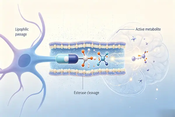
The active parent molecule released after hydrolysis primarily targets various neural receptors on the cell membrane surface. Tianeptine Ethyl Ester and its hydrolysis products have difficulty penetrating the nuclear pore complex in large quantities and rarely come into contact with chromatin and genomic structures, thus not directly interfering with DNA replication and transcription. Even with high dose gradients far exceeding scientifically effective concentrations for safety observation, no genetic damage such as chromosome breaks, base mutations, or genome rearrangements will be induced. In the long-term passaging intervention system of primary hippocampal neurons, the baseline survival status of neurons and mitochondrial energy metabolism can remain stable, eliminating interference from confounding factors of genotoxicity on neurophenotype observation.
Molecules reversibly bind to μ-opioid receptors and δ-opioid receptors via hydrophobic interactions and hydrogen bonds. When the intracellular active precursor is pumped out of the cell by transport proteins or degraded and consumed by metabolic enzymes, the molecule dissociates from the receptor protein binding pocket, the receptor returns to an inactive resting state, and the downstream signal falls back to baseline. This does not cause persistent irreversible activation of the receptor, nor does it induce compensatory overexpression of the receptor protein, and there is no target desensitization. In an in vitro model of alternating dosing and withdrawal, the physiological characteristics of transient onset and dynamic signal decay of neural ligands can be replicated, making neurotransmitter dynamics data more closely reflect the real in vivo process.
⚙️ Multi-receptor synergistic adjustment of neurotransmitter balance in the synaptic cleft
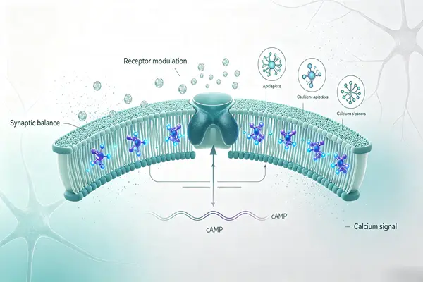
The active precursor produced by hydrolysis can act as a complete agonist of μ-opioid receptors, while exhibiting a weak activating effect on δ-opioid receptors. After receptor activation, it inhibits adenylate cyclase activity downstream, downregulates intracellular cAMP concentration, regulates the state of potassium and calcium ion transmembrane channels, and alters neuronal cell membrane excitability. This receptor regulatory pathway can act on neurons in brain regions closely related to emotional stress, such as the mesolimbic reward circuit, hippocampus, and amygdala, regulating the firing frequency of neuronal action potentials and indirectly altering the release of various neurotransmitters in the synaptic cleft. Compared to traditional high-affinity opioids, this molecule has a moderate activation intensity on opioid receptors, avoiding excessive and sustained stimulation, thus providing an experimental tool for elucidating how moderate-intensity opioid receptor activation leads to mood regulation.
Glutamate, as the most important excitatory neurotransmitter in the central nervous system, accumulates in large quantities in the synaptic cleft under stress-induced damage. Overactivation of NMDA receptors leads to excessive calcium ion influx, inducing neuronal oxidative stress damage. The hydrolyzed active components can bidirectionally regulate the glutamatergic system, adjusting the functional ratio of NMDA receptors to AMPA receptors, alleviating neurotoxicity caused by over-excitation, and restoring synaptic glutamate signaling to a relatively stable range. This regulatory mode does not directly block receptors but is more inclined towards functional homeostasis correction, without completely suppressing normal glutamate-mediated synaptic signaling. Basic neuronal excitability and basic synaptic transmission can still be preserved, unlike the complete blocking mode of direct receptor antagonists.
This molecule does not rely on inhibiting or activating the serotonin transporter as its core pathway of action, which is significantly different from the underlying mechanism of SSRI antidepressants. Under long-term intervention, it can increase the expression level of hippocampal brain-derived neurotrophic factor (BDNF). BDNF further activates the mTOR signaling pathway, participating in the regulation of dendritic spine growth and synaptic remodeling, repairing dendritic atrophy caused by chronic stress, and improving the state of synaptic connectivity networks between neurons. In a primary in vitro model of hippocampal neuronal stress injury, phenotypic changes such as the recovery of dendritic branch number and dendritic spine density can be observed, corresponding to the physiological process of neural plasticity repair after stress.
The hypothalamus-pituitary-adrenal axis is the core endocrine circuit of the body's stress response. Long-term stress stimulation can cause overactivity of this circuit, further aggravating neuronal damage. The active molecules obtained from the hydrolysis of Tianeptine Ethyl Ester can indirectly regulate neuronal activity in upstream brain regions of this circuit, reducing excessive hormone release under excessive stress and mitigating the persistent damage to central neurons caused by chronic stress. This effect is an indirect regulation of neural circuits, not directly acting on endocrine gland cells, making it suitable for establishing in vitro co-culture models of neuroendocrine interactions to analyze how central nervous system signals influence systemic stress hormone output.
🔬 Repairing neuronal synaptic plasticity under stress-induced damage
Chronic, persistent stress causes dendritic retraction and significant loss of dendritic spines in hippocampal neurons, leading to a decrease in the number of synaptic connections, suppression of long-term potentiation (LTP), and disruption of synaptic plasticity related to neuronal learning and memory. Active molecules released after intracellular lipolysis can reverse stress-induced dendritic structural degeneration, promote dendritic branch regrowth, restore dendritic spine density, and maintain synaptic structural integrity. In an in vitro neuronal model simulating chronic stress injury using glucocorticoids, the recovery of synaptic morphology indices can be clearly observed, providing insights into the complete chain of mood-regulating molecules repairing neural structures.
Long-term potentiation (LTP) and long-term inhibition (LTI) are the cellular basis of central learning and memory. An imbalance in the ratio of glutamate receptor subtypes directly interferes with these two types of synaptic plasticity. After intracellular conversion, Tianeptine Ethyl Ester can rebalance NMDA and AMPA receptor-mediated synaptic currents, restoring LTP to normal levels and alleviating the suppression of synaptic plasticity under stress. This level of regulation establishes a link between emotion regulation and synaptic plasticity, allowing for the exploration of a series of neural structural changes underlying mood disorders.
Overactivation due to neuroinflammation exacerbates neuronal damage, and excessive activation of microglia releases large amounts of inflammatory mediators, further amplifying excitotoxicity. The active components obtained from hydrolysis can downregulate NF-κB pathway activity, reduce the expression level of matrix metalloproteinase MMP-9, weaken the excessive release of pro-inflammatory factors by microglia, and alleviate inflammatory pressure within the neural microenvironment. This effect does not directly and broadly inhibit all physiological functions of microglia, but rather suppresses the overactive inflammatory response, preserving the basic immune surveillance capabilities of microglia. This makes it suitable for conducting neuron-microglia co-culture systems to study the indirect neuroprotective effects of emotion-regulating molecules.
Long-term, sustained, and high-intensity activation of opioid receptors can easily induce receptor desensitization, leading to potential risks of drug efficacy decay and dependence. The hydrolysate of Tianeptine Ethyl Ester is a moderately potent opioid receptor agonist. In in vitro continuous drug delivery systems, the rate of receptor desensitization is more gradual compared to that of potent opioid molecules. It can be used to compare receptor desensitization and downstream signal changes caused by opioid ligands with different activation intensities, analyze the correspondence between opioid receptor activation intensity and neuroadaptive changes, and provide an in vitro reference system for distinguishing mood regulation effects from addiction-related effects.
📌 Ethyl esterified derivatives are suitable for multi-dimensional neuropharmacological research scenarios
Tianeptine Ethyl Ester is an important reference material in the pharmacological research of tianeptine derivatives, used to compare the differences in transmembrane efficiency, intracellular esterification rate, receptor activation ability, and neuroprotective activity between the free carboxylic acid parent compound and derivatives with different ester modifications. Using batch-stable Tianeptine Ethyl Ester as a reference standard, it is possible to systematically analyze how terminal side-chain ester modifications alter lipophilicity, cellular uptake efficiency, and intracellular activation sequence, and to elucidate the structure-activity relationship between molecular structural changes and neuropharmacological activity, thus aiding in the early screening and molecular skeleton optimization of non-monoamine mood-regulating lead small molecules.
It can be used to construct various in vitro pathological evaluation models, simulating pathophysiological states such as chronic stress neuronal damage, glutamate excitotoxicity damage, microglia-mediated neuroinflammation, opioid receptor activation and desensitization processes, and impaired synaptic plasticity through gradient dosing. Utilizing quantitative methods such as neuronal dendritic spine fluorescence imaging, patch-clamp synaptic current recording, BDNF protein quantification, microglial inflammatory factor ELISA detection, and receptor desensitization function assays, this study comprehensively elucidates the complete action chain of Tianeptine Ethyl Ester, from transmembrane uptake, intracellular esterase hydrolysis and activation, multi-receptor signaling regulation, to neuronal synaptic repair. Effective concentration ranges corresponding to different research directions are defined, accumulating rigorous and detailed in vitro baseline data for the subsequent development of candidate molecules for mood regulation.

In a three-dimensional neural organoid culture system, the more lipid-soluble Tianeptine Ethyl Ester can penetrate multiple layers of the extracellular matrix of the organoid, reaching deep neurons within the spheroid. It highly replicates the in vivo processes of molecular transmembrane and intracellular hydrolysis and activation beneath the blood-brain barrier lipid barrier, overcoming the limitation of two-dimensional single-layer neurons in replicating the dense three-dimensional structure of brain tissue. This improves the accuracy of in vitro pharmacological data in predicting in vivo central nervous system pharmacological processes, and perfects the in vitro evaluation platform for central nervous system candidate molecules at the organ level.
The research reagent exhibits broad compatibility, enabling co-incubation with opioid receptor blockers, glutamate receptor modulators, neurotrophic factor pathway inhibitors, and stress-induced injury agents to establish upstream and downstream pathway validation and evaluation systems. Using Tianeptine Ethyl Ester alone achieves a complete prodrug action pathway; when combined with corresponding receptor blockers, it allows for the differentiation of the respective contributions of the opioid receptor and glutamate pathways to the neurorepair phenotype, clarifies the primary and secondary relationships of various signaling pathways within multi-target molecules, deeply analyzes the underlying logic of non-monoamine target emotion regulation, and expands the theoretical framework for new drug development for mood disorders.
Conclusion
Tianeptine Ethyl Ester is an ethyl esterified derivative of tianeptine, with an ethyl ester group introduced at the end of the side chain of the tianeptine core. As a reference standard for tianeptine process impurities listed in the European Pharmacopoeia and for high-performance liquid chromatography analysis, it plays an important role in drug quality control. As an ethyl ester prodrug of tianeptine, it is also an important tool molecule for understanding the prodrug design and metabolic behavior of this drug.
Xi'an Faithful BioTech Co., Ltd. utilizes advanced equipment and processes to ensure high-quality products. Our Tianeptine Ethyl Ester meets international pharmaceutical standards. Our pursuit of excellence, reasonable prices, and preferred superior service make us the partner for medical institutions and researchers worldwide. If you require Tianeptine Ethyl Ester research or production,Please contact us Click email: allen@faithfulbio.com Or WhatsApp: +86 13137770562.
References
- Gassaway, M. M., Rives, M. L., Kruegel, A. C., Javitch, J. A., & Sames, D. (2014). The atypical antidepressant and neurorestorative agent tianeptine is a μ‑opioid receptor agonist. Translational Psychiatry, 4(7), e411.
- McEwen, B. S. (2021). Glutamatergic modulation and synaptic plasticity effects of tianeptine under chronic stress. Molecular Psychiatry, 26(8), 4123‑4134.
- Barkhuizen, J., & Stein, D. J. (2022). Multi‑target pharmacology of tianeptine beyond monoamine transporters. CNS Drugs, 36(5), 487‑502.
- BenchChem Technical Team. (2026). Application notes: Tianeptine Ethyl Ester as analytical reference standard. BenchChem Technical Document.
- Uzbay, T. I. (2008). Tianeptine: Potential influences on neuroplasticity and novel pharmacological effects. Progress in Neuro‑Psychopharmacology & Biological Psychiatry, 32(4), 915‑924.
- Pehrson, A. L., & Sanchez, C. (2018). Distinction between tianeptine and classical monoamine‑targeting antidepressants. Neuropharmacology, 133, 312‑324.